Showing posts with label article. Show all posts
Stupid act by UMers
7/03/2009 01:21:00 AM | Author: fadzly
1. The title was a bit harsh.

2. I'm actually talking about one of University Malaya (UM) student from Academy of Islamic Studies (API) which is also the President of PMIUM currently doing stupid act.

3. I'm not talking about politics. I'm not a politician, and i hate politics thus, there is no way that I'm gonna talk politics here.

4. Because I'm the most seniors undergrad stud in UM currently, it's important for me to address this messages to him. I care my junior, is that a problem? My junior have a duty and responsible to their parents.

5. Firstly, for me his act is certainly not an intellectual move which is supposedly to be expected from every student from UM.

6. We are not a politician, not an extremist, not a another 'RPK'. We're only a 'Mahasiswa Biasa'.

7. You might seem big to us because you're President of PMI, but do you think you are big enough out here?

8. Do you think that you big enough you can do anything against the law and because of that you run from getting caught from the cops?

9. You're the 'role model' of all the student in UM. Do you think this act should be done on every students here?

10. Or you just want to have a 'publisiti murahan' so that all Malaysians know you.

11. Don't make assumption, where u based only on youtube video without any fact.

12. Don't make a statement threaten to the university admins of having a riot. If you do, i do agree if AUKU is implemented on you.

13. I suggest you shouldn't be doing it. Better for you to do good deeds and welfare to the students.

14. Where is your 'kurangkan yuran universiti' and also 'Riba PTPTN' fight anyway? My yuran is so expensive, and yet i don't have any $$ left in my pocket!

15. University can throw you away anytime they want, because you have done the DUMBESS act which is against the law.

16. And yet they have not done it because they think that you're the Leaders of Islamic Student? Of course not, because the investigation have not been concluded and you've not been prove to be the culprit who behind this DS Rosmah C4 wall painting.

17. If DS Rosmah was really doing all those nasty things, dont you think by putting an oil/petrol on the carpet and try to harm her by putting up a fire are the best way to punish her?

18. Do you think you wont be having a fair trials? Do you think that Anwar Ibrahim and RPK doesn't have a fair trial? Do you think that you'll be beaten at the eye and having a 'panda eye' like Anwar Ibrahim in the lockups?

19. If you have all this you might having a delusion of persecutory which might indicate you a schizophrenic. If it does, you should meet me, here, in Psychiatric ward in UMMC.

20. Maybe, i mean it, maybe, you might or will be given an exemption from the trial.

21. So please don't ask your friends to become one like you.

22. Don't you think that by AUKU is important now?
read more about“Stupid act by UMers”
We've got our 1st Influenza A transmission Man to Man
6/17/2009 05:30:00 PM | Author: fadzly
Hey, first of all i don't know it's real or not because it's from Malaysiakini.com. But if this is real, this would be the very bad. Why? Because this patient was send to UMMC!!!!!!!!
Damn I don't want to go to the ward after this!! An irresponsible act by a 17 year old girl who don't contact to the ministry after getting exposure to patient of the 12th case of Influenza A in Malaysia. This would be the 19th case and just now, the 20th case has just been reported! Because of this, no wonder WHO has declaring pandemic, the Influenza A gene has evolve from swine to human transmission, to human to human transmission! Good thing that there is no death case reported here. And when there is death case, it would be very very bad to our country.

Updated: Meanwhile our cases have reached 23rd cases with 5 new cases today according to the MOH here.
Nevermind. The patient has been transferred to HKL already.. My bad.. No excuse not to go to the ward..


kemaskini 4.59pm Jangkitan selesema A (H1N1) semakin serius di negara ini, susulan berlakunya penularan wabak tersebut di peringkat tempatan yang pertama pada pesakit ke-19 daripada 23 kes, sejak sebulan lalu.

Pesakit berkenaan, seorang pelajar perempuan 17 tahun, dijangkiti daripada pesakit ke-12 akibat kegagalan kuarantin apabila tidak memaklumkan pernah berhubung dengan pelajar perubatan kes terdahulu.

Akibatnya, menurut satu kenyataan Kementerian Kesihatan hari ini, kegagalan itu menyebabkan 20 orang lagi berkemungkinan terdedah kepada jangkitan H1N1.

Kementerian juga memaklumkan bahawa lima lagi kes influenza itu dikesan dalam tempoh 24 jam yang lalu.

"Salah satu dari kes yang disahkan positif Influenza A (H1N1) adalah kes penularan tempatan yang pertama berlaku di negara ini," kata Ketua Pengarah kesihatan, Tan Sri Dr Mohd Ismail Merican.

"Kes pertama penularan tempatan (kes ke-19) adalah seorang pelajar perempuan berumur 17 tahun, warganegara Malaysia yang tidak mempunyai sejarah melawat negara (asing) yang telah diisytiharkan berlaku wabak Influenza A(H1N1).

"Beliau telah mula menunjukkan simptom batuk dan selesema pada 14 Jun 2009 jam 9 malam, dan mula demam pada keesokan harinya. Pada pagi 15 Jun 2009, kes telah pergi ke Pusat Perubatan Universiti Malaya seterusnya dirujuk ke Hospital Sungai Buloh jam 10.00 pagi.

"Semasa penyiasatan, kes ke-19 melaporkan bahawa beliau adalah kontak kepada kes ke-12 iaitu pelajar perubatan yang telah disahkan positif Influenza A(H1N1) pada 13 Jun 2009.

"Namun sebelum ini beliau tidak pernah diisytiharkan sebagai salah seorang kontak kepada kes ke-12.

"Ini menyebabkan pengawasan dan kuarantin awal ke atas kes ke-19 ini gagal dilakukan dan menyebabkan 20 orang lagi (kontak) terdedah kepada jangkitan influenza A(H1N1)."

Dr Mohd Ismail menambah, sangat penting semua kes yang disahkan
positif H1N1 memberikan maklumat lengkap supaya pengawasan dan kuarantin awal dapat dilakukan kepada semua orang yang pernah berhubung dengan mangsa, seterusnya mengelakkan risiko penularan kepada orang lain.

"Tindakan undang-undang bawah Akta Pencegahan dan Pengawalan Penyakit Berjangkit 1988 boleh diambil sekiranya mereka gagal memberikan maklumat yang lengkap," katanya.

Tambahnya lagi, sesuai dengan keputusan Mesyuarat Jawatankuasa Teknikal Kejadian Influenza A(H1N1) bil. 5/2009, kementerian juga mengeluarkan arahan seperti berikut:

bullet buttonOrang awam tidak digalakkan untuk melawat hospital. Kanak-kanak berumur bawah 12 tahun dilarang pergi atau dibawa melawat ke hospital kecuali untuk mendapatkan rawatan.

Setiap pesakit hanya dibenarkan menerima dua orang pelawat berumur lebih 12 tahun pada satu-satu masa. Masa lawatan di hospital kerajaan dan swasta akan dipendekkan.

bullet buttonSaringan demam akan dilakukan kepada semua pelawat yang
datang melawat pesakit di semua hospital kerajaan dan swasta.

Kementerian Kesihatan juga memohon kerjasama kepada penumpang, anak kapal dan kontak kepada kes agar menghubungi melalui talian 03-88 81 02 00 atau 03-88 81 03 00 untuk mendapatkan nasihat kesihatan termasuk keperluan kuarantin di rumah.
read more about“We've got our 1st Influenza A transmission Man to Man”
Type and Indication of Blood Transfusion
6/13/2009 09:15:00 PM | Author: fadzly
The second short note question would be about blood transfusion. Safe blood products, used correctly, can be life-saving. However, even where quality standards are very high, transfusion carries some risks. If standards are poor or inconsistent, transfusion may be extremely risky. No blood or blood product should be administered unless all nationally required tests have been carried out.


Blood component:
1 A constituent of blood, separated from whole blood, such as:
  • Red cell concentrate
  • Red cell suspension
  • Plasma
  • Platelet concentrates
2 Plasma or platelets
3 Cryoprecipitate, prepared from fresh frozen plasma: rich in Factor VIII and fibrinogen

Plasma derivative
Human plasma proteins prepared under pharmaceutical manufacturing conditions, such as:
  • Albumin
  • Coagulation factor concentrates
  • Immunoglobulins


WHOLE BLOOD
  • 450 ml donor blood
  • 63 ml anticoagulant-preservative solution
  • Haemoglobin approximately 12 g/ml
  • Haematocrit 35%–45%
  • No functional platelets
  • No labile coagulation factors (V and VIII)
Indication
  • Red cell replacement in acute blood loss with hypovolaemia
  • Exchange transfusion
  • Patients needing red cell transfusions where red cell concentrates or suspensions are not available


RED CELL CONCENTRATE

  • 150–200 ml red cells from which most of the plasma has been removed
  • Haemoglobin approximately 20 g/100 ml (not less than 45 g per unit)
  • Haematocrit 55%–75%
Indication
  • Replacement of red cells in anaemic patients
  • Use with crystalloid replacement fluids or colloid solution in acute blood loss

LEUCOCYTE-DEPLETED RED CELLS

  • A red cell suspension or concentrate containing <5>
  • Haemoglobin concentration and haematocrit depend on whether the product is whole blood, red cell concentrate or red cell suspension
  • Leucocyte depletion significantly reduces the risk of transmission of cytomegalovirus (CMV)
Indication
  • Minimizes white cell immunization in patients receiving repeated transfusions but, to achieve this, all blood components given to the patient must be leucocyte-depleted
  • Reduces risk of CMV transmission in special situations
  • Patients who have experienced two or more previous febrile reactions to red cell transfusion


PLATELET CONCENTRATES

Single donor unit in a volume of 50–60 ml of plasma should contain:
  • At least 55 x 109 platelets
  • <1.2>
  • <0.12>
Indications
  • Treatment of bleeding due to:
  • — Thrombocytopenia
  • — Platelet function defects
  • Prevention of bleeding due to thrombocytopenia, suchas in bone marrow failure


FRESH FROZEN PLASMA

  • Pack containing the plasma separated from one whole blood donation within 6 hours of collection and then rapidly frozen to –25°C or colder
  • Contains normal plasma levels of stable clotting factors, albumin and immunoglobulin
  • Factor VIII level at least 70% of normal fresh plasma level
Indications
  • Replacement of multiple coagulation factor deficiencies: e.g.
  • — Liver disease
  • — Warfarin (anticoagulant) overdose
  • — Depletion of coagulation factors in patients receiving large volume transfusions
  • Disseminated intravascular coagulation (DIC)
  • Thrombotic thrombocytopenic purpura (TTP)

CRYOPRECIPITATE

  • Prepared from fresh frozen plasma by collecting the precipitate formed during controlled thawing at +4°C and resuspending it in 10–20 ml plasma
  • Contains about half of the Factor VIII and fibrinogen in the donated whole blood: e.g. Factor VIII: 80–100 iu/ pack; fibrinogen: 150–300 mg/pack
Indication
* As an alternative to Factor VIII concentrate in the treatment of inherited deficiencies of:
— von Willebrand Factor (von Willebrand’s disease)
— Factor VIII (haemophilia A)
— Factor XIII
* As a source of fibrinogen in acquired coagulopathies:
e.g. disseminated intravascular coagulation (DIC)


HUMAN ALBUMIN SOLUTIONS

Prepared by fractionation of large pools of donated plasma

Indication
  • Replacement fluid in therapeutic plasma exchange: use albumin 5%
  • Treatment of diuretic-resistant oedema in hypoproteinaemic patients: e.g. nephrotic syndrome
  • or ascites. Use albumin 20% with a diuretic
  • Although 5% human albumin is currently licensed for a wide range of indications (e.g. volume replacement, burns and hypoalbuminaemia), there is no evidence that it is superior to saline solution or other crystalloid replacement fluids for acute plasma volume replacement

COAGULATION FACTORS


Factor VIII concentrate
  • Treatment of haemophilia A
  • Treatment of von Willebrand’s disease: use only preparations that contain von Willebrand Factor

PLASMA DERIVATIVES CONTAINING FACTOR IX

Prothrombin complex concentrate (PCC)
Factor IX concentrate
  • Treatment of haemophilia B (Christmas disease)
  • Immediate correction of prolonged prothrombin time


read more about“Type and Indication of Blood Transfusion”
POSTOPERATIVE MANAGEMENT
6/12/2009 04:49:00 PM | Author: fadzly
End of Posting Written Test
Lolz.. This morning short note test come out to be complication of operation.. Damn i don't like this kinda question. I hate short notes.. really.. But nway, i've made an article for us to look what we've answer and filling all the blanks that we have.. But hey, this is not an answer scheme or what so ever. It just for our understanding and merely a revision before we get ourself into a House Officer..

The role of the junior doctor postoperatively is to check that the patient has recovered from the anaesthetic, look at their observation charts and check their fluid balance. The operation note should have a section on specific postoperative management written by the surgeon, and is a guide that should be followed. For example, following a vascular graft operation — say, to the leg — you should always check the pulses, capillary refill and toe movement in the involved leg to ensure that the graft has not blocked off. A common question for exams concerns complications of surgery.

Complications
All operations carry a risk of complications. These can be divided into general and specific.

* General complications include those pertaining to the anaesthesia itself and those that can occur after any operation, such as a chest infection or DVT.
* Specific complications are those that occur because of the individual operation itself, such as cutting a nerve.

You can subdivide this classification by time, into complications that occur immediately, within the first 24 h; early, within the first week or so; late postoperative, occurring within the first month or so; and long term.

General immediate complications include those due to the anaesthetic, such as direct trauma to the mouth when intubating and reactions to the anaesthetic (inherited disorders or idiosyncratic reactions).

Other early complications including
* Chest infections
* Urinary retention or infections
* Deep vein thrombosis
* Bed sores.

Specific complications depend on the nature of the operation. In this category haemorrhage and wound infection are important.

Haemorrhage
This can be divided into primary, reactionary and secondary haemorrhage.

* Primary haemorrhage occurs during the operation, when a vessel is cut.
* Reactionary haemorrhage is when at the end of the operation the wound looks dry, but when the patient’s blood pressure and cardiac output rise to normal levels, bleeding begins, presumably from vessels that were not properly ligated during the operation.
* Secondary haemorrhage, occurring several days after the operation, is usually attributed to infection that erodes through a vessel.

Wound Infections
These are most commonly caused by S. aureus (increasingly MRSA), although coliforms such as E. coli are also important. Wound infection is more likely if

• The operation is dirty (e.g. abdominal surgery)
• The duration of the operation is long (greater than 2 h)
• The patient is more susceptible (e.g. old age, immunosuppression, diabetes)

Minor wound infections, with a little redness and slight discharge, are relatively common and usually need just simple measures, such as regular wound dressing and perhaps antibiotics. More severe infections, common after abdominal operations, usually occur in the first week or so. The wound looks inflamed, and there may be cellulitis, discharge or localised abscess formation. The wound should be swabbed and maybe antibiotics started, but the only correct treatment for an abscess is drainage. This may mean simply removing a few of the surgical clips, and probing the wound, allowing the pus to discharge, or a further surgical procedure to open up the wound. The wound is then left to heal by secondary intention (i.e. to heal itself from within, with no further suturing).

Wound Dehiscence
This is an uncommon problem. It is usually due to an inadequate repair of the tissues (but infection, poor blood supply, malnutrition and steroids may all play a part in poor wound healing). Dehiscence usually occurs about a week after the operation. A warning sign is a serosanguinous discharge from the wound a few days before. The wound suddenly bursts open and in the case of a laparotomy the bowel protrudes outwards and is extremely alarming for the patient and the nursing staff. Sterile soaked swabs should be placed over the wound and the patient taken back to the theatre for repair.

serasenguinous discharge

An example of general and specific complications pertaining to a - gastrectomy is outlined below.

Click to enlarge

The commonest reason that a junior doctor gets called to the ward is to write up fluids or to see a patient with postoperative pyrexia or poor urine output.

POSTOPERATIVE PYREXIA
A small rise in temperature is common postoperatively. If the temperature spikes above 38'C or persists, then you should consider and look for the seven C's as potential causes. This is a common viva question.

1. Chest. Chest infection
2. Catheter. UTI
3. CVP line. Infected
4. Cannula. Superficial thrombophlebitis (solved by removing the cannula)
5. Cut. Wound infection
6. Collection. Subphrenic or pelvic abscess (may indicate a failure of anastomosis)
7. Calves. DVT (rumbling pyrexia in second postoperative week)

A chest infection is very common postoperatively, especially in patients who smoke or have pre-existing poor respiratory function. The mucus secretions are not cleared; these then clog up the smaller bronchi, which leads to collapse of the air spaces distal to the blockage (atelectasis). Inhaled organisms then infect the collapsed segments. In addition, thoracic and upper abdominal incisions cause pain and stop the patients from coughing up the secretions, and so they are much more likely to have basal atelectasis and develop chest infections. These patients should therefore be given adequate analgesia, have vigorous physiotherapy, and be encouraged
to cough up the phlegm (ideally whilst holding their wounds — applicable for chest and abdominal wounds).

Abscesses
A deep collection, such as a subphrenic or pelvic abscess, can occur after the patient has had generalised peritonitis. The patient usually presents with general malaise, nausea, pain (a subphrenic abscess may also cause pain felt in the shoulder tip), a swinging pyrexia and localised peritonitis.

A pelvic abscess often occurs 4–10 days postop., whereas a subphrenic abscess usually occurs a bit later, 7–21 days postop. Clinically, the patient appears to be recovering well, but then develops a fever and starts to feel unwell. The white cell count may be raised and a collection is identified on ultrasound or CT. Treatment is by drainage, either percutaneously under ultrasound or CT guidance, or by an open procedure. A drain is usually left in situ.

Anastamotic leak
A small anastomotic leak usually causes a localised abscess which becomes sealed off by the omentum and the bowel. Clinically, the patient is slow to recover, but usually improves with intravenous antibiotics and fluids and delayed return to food. A larger anastomotic breakdown causes the patient to be very unwell, with anything from local peritonitis through to a rigid abdomen and septicaemia. The abscess needs to be drained, the peritoneal cavity washed out, and the two ends of the failed anastomosis can be brought out as temporary stomas.

Thrombosis
A diagnosis of DVT and pulmonary embolus (PE) in the first instance is essentially a clinical one, as treatment is usually instituted before definitive diagnosis is made. A PE usually presents with pleuritic chest pain (stabbing and worse on inspiration). The textbooks tend to describe the findings you would see in a massive PE, although more commonly in the smaller PEs the findings are less impressive. Usually, the patient is tachycardic, maybe with a low-grade fever and maybe tachypnoeic, but not much else and they may even be asymptomatic. The ECG usually shows sinus tachycardia (the classic S1Q3T3, which most students know about, occurs when there is a large amount of right heart strain, in a large PE, and is rarely seen). The CXR is usually unhelpful but may show a small area of linear atelectasis.

S1Q3T3

Blood gas analysis is essential and you would expect to find a low PO2 (due to ventilation/perfusion mismatches) and a low PCO2 due to hyperventilation. Examination of the calves may or may not reveal evidence of a DVT. If a DVT or PE is suspected, a heparin infusion can be started before investigation but always check with a senior colleague before doing that, especially if the patient has had recent surgery.

To diagnose a DVT you can use duplex ultrasound (or a venogram). To diagnose a PE you can request a ventilation–perfusion scan, although the gold-standard is pulmonary angiography (note nowadays a spiral CT is used in some centres).

Other less common causes for a fever include infective diarrhoeas, drug reactions and blood transfusion reactions. If faced with a patient with a pyrexia you would obviously find out a little history and examine the patient properly. In a long case exam situation you could answer along the lines of, ‘I would listen to the chest, examine the abdomen, check the cannula sites, inspect the wound, etc. … My investigations would depend on my clinical findings but may involve sending a urine specimen, a full blood count and blood cultures, sending wound swabs or the tip of the central line for culture, etc.’

DRAINS

Collections within a wound (especially if they contain blood) are the perfect medium for colonisation of bacteria and hence infection.

A drain can be used to remove anticipated collections within a wound, but should never be used as a substitute for adequate haemostasis at the time of surgery.

Drains can be closed or open:

Closed drainage includes suction drainage (e.g. Redivac) where the collection is attracted into a container either by gravity or suction. This can then potentially reduce the risk of infection when used for large spaces or cavities, such as after a mastectomy or joint replacement. Drains are usually removed as soon as possible (usually 24–48 h) or as soon as the losses begin to tail off. Drains can also introduce infections and so they should not be left in for any longer than needed.

Open drainage (e.g. a piece of corrugated tubing with one end in the wound and the other in the dressing), allows small losses to escape from the wound. This is often employed in established abscesses after incision and drainage to allow any remaining collection a passage out of the wound. Some surgeons like to withdraw this type of drain in stages to allow the track to collapse behind it.

Other drains commonly asked about in exams include chest drains, T-tubes and percutaneous nephrostomies.

POSTOPERATIVE POOR URINE OUTPUT
This is a common exam question and can be classified as prerenal, renal or postrenal. The commonest causes of failure to pass urine postoperatively are postrenal.

Postrenal problems (commoner in males) include obstruction caused by a large prostate or a blocked catheter. Also, the patient may find initiation of micturition difficult for the following reasons:

1. Anticholinergic drugs or those with alpha adrenergic effects (e.g. the anaesthetic)
2. Pain (e.g. after a hernia repair)
3. Inhibition (e.g. because of strange surroundings or a nurse continually asking them if they have passed urine)
4. Opiates or epidural anaesthetics

Once the bladder reaches a certain volume of distension it fails to function properly and the patient goes into retention. Benign prostatic hypertrophy is an important predisposition and these patients are more likely to go into retention.

Prerenal causes are due to renal hypoperfusion because of either hypovolaemia or heart failure.

Renal causes — Acute renal failure is usually due to acute tubular necrosis.

How to handle?
Junior doctors are commonly called to see patients who have failed to pass urine postoperatively. Often this will be a patient you are covering but have not met before. It is, therefore, worth spending a little time getting a history and reading the patient’s notes. You should find out the type and date of operation and also search for clues pointing to whether the problem is prerenal or postrenal. Ask if there is any pain; an enlarged bladder causes suprapubic pain, although this is difficult to differentiate from pain in an abdominal wound (one exception is when there is an epidural in situ and there is no pain).

On examination you should look for signs of hypovolaemia (dry mucous membranes, decreased skin turgor, tachycardia, etc.), signs of heart failure (shortness of breath, tachycardia, raised CVP or JVP and bibasilar crepitations, peripheral oedema, etc.). A distended bladder palpable just above the pubis is dull to percussion and usually tender, making the patient want to pass urine, when compressed.

You can initially try conservative measures such as analgesia, privacy, sitting in a warm bath, etc., but if that fails then catheterisation is indicated. If you suspect that the cause is postrenal (i.e., distended bladder and discomfort), then the diagnosis is proven by catheterisation. A large residual volume of urine should drain (usually about 500 ml or more).

If the patient already has a catheter in situ, then the catheter should be flushed to ensure it is not blocked. If the urine coming out of the catheter is small amounts of concentrated dark urine, then the cause is likely to be prerenal anyway. You could dipstick the urine, testing for a high specific gravity (more than 1020) to prove this.

You should check the fluid balance charts (IO chart). Observe how much fluid has gone in before, during and since the operation and the measured urine output. Remember that a long laparotomy can lead to large losses of fluid by evaporation and this will not be measured on the charts. A urine output of less than about 30 ml/h is poor (oliguria). The patient should be catheterised anyway at this point to measure the hourly urine output.

U & Es should be sent; if there is a prerenal cause, the urea will be raised. If from your clinical examination and your assessment of the fluid balance charts you think the patient has a prerenal cause, then you should try a fluid challenge.

Fluid Challenge — 250–500 ml of normal saline given as a stat dose (unless you suspect blood loss as the cause, in which case the patient may be shocked and should be given colloids and blood). Then, observe the urine output over the next hour. If the urine output picks up, you have shown the patient to be in need of more fluid and the next bag should be speeded up.

If the patient has heart failure, the urine output will not pick up and the patient may become a little more breathless and the CVP might rise. If this occurs, a bolus dose of a loop diuretic, such as 40 mg of frusemide, will lead to a diuresis and a fall in the CVP. If the patient does not have a central line in at this point, you should ask a senior colleague for advice, as a central line is really necessary for knowing exactly what the state of vascular filling is and will help in the management.

Only after you have excluded a postrenal and a prerenal cause can you assume that there may be a renal problem and acute tubular necrosis has occurred. The creatinine will be raised and you should measure the urine and plasma osmolalities. The ratio of urine to plasma osmolality will be less than 1 (as opposed to prerenal oliguria, where the ratio will be more than 1). Advice from a renal physician should be sought early.

That's all folks.. Hope can help you a bit. I need a great night sleep tonight after tired using my brain for 12 hours straight. Good night.

read more about“POSTOPERATIVE MANAGEMENT”
PRE-OPERATIVE MANAGEMENT
6/05/2009 08:11:00 PM | Author: fadzly
I'm wondering to have an article about what a House Officer should do when they in the surgery posting. I hope this helpful for my 'old classmates' who are going to have and induksi this week. Congrates to them they are going to start their work! When you are a surgical house officer (or Formula One), part of your role preoperatively will be to clerk the patients and prepare them for the theatre or their investigations.

A clerking consists of the history of the presenting complaint, past medical history, drug history, family history and social history. You should then examine the patient fully, looking first at their general health and whether they are fit enough for the operation, and if not you should be thinking of ways to optimise their health, such as using preoperative nebulisers for an asthmatic. The clerking also allows other problems to be picked up.

If, when the patient arrives on the ward, you feel that the diagnosis made in the out-patient clinic has changed, you should inform a senior colleague before the operation is booked. For example, if a patient was admitted for an excisional biopsy of a lymph node that has completely disappeared when you examine them, you should inform your consultant, as the operation might need to be cancelled.

In the main, appropriate investigations should be performed before surgery and this is often a good question for a viva examination. For example, before a laparascopic cholecystectomy is performed the patient should have had an ultrasound to confirm the presence of gallstones, and a set of liver function tests. The house officer should discuss the order of patients on the operating list with the operating surgeon. Usually, children are placed first on the list, as this is nicer for the child and the parents; also they find it hard to go without food for long periods. If special equipment is needed in the theatre, such as the image intensifier for X-rays or laparascopic equipment, then these should be discussed with the theatre staff (and radiographers) the day before.

Specialist nurses who have expertise in certain areas such as breast disease, wound management and stomas should be involved preoperatively in all appropriate cases. For example, a patient who is likely to need a colostomy or ileostomy should be seen by the stoma nurse specialist several days before the operation. This allows for patient education (i.e. answering of any questions and worries), and also the site where the stoma will be cited is marked (note that the patient should be standing, so
as to position it in the most appropriate place).


Comorbidity

Many patients have problems other than the one that is being operated upon. These may be social and as such need a social work or occupational therapy referral. For example, if the patient has difficulty climbing stairs and there is no lift, then there may be a need to arrange for a stair lift or rehouse into ground floor accommodation.

The patients may have intercurrent medical problems such as diabetes, hypertension or chronic obstructive pulmonary disease (COPD). They may also be on drugs such as steroids or anticoagulants. When clerking the patient you should be looking out for these, and if you think they may affect the operation, then you should inform the anaesthetist or the consultant in charge of the patient.

As a house officer the tests you need to consider preoperatively include blood tests, such as a full blood count, a sickle screen if at risk (this includes anyone of Afro-Caribbean origin), and either a group and save or a cross-match. You should X-match any patient at risk of blood loss extensive enough to need replacing — for example, the anticipated blood loss from an anterior resection is about two units, but to be safe we usually X-match four units. The blood is kept in the refrigerator ready for use. If it is not used it goes back to the blood bank for storage.

A young healthy person, in general, requires no preoperative investigations, but if at all unsure then you should ask the anaesthetist what they would like performed (for example, some anaesthetists like to have a recent full blood count on all females of childbearing age). The National Institute for Clinical Excellence (NICE) have issued guidelines on preoperative
investigation available here. If the patient is hypertensive or on diuretics then a U & E (urea, creatinine and electrolytes) to assess renal function must be performed. An ECG is necessary on anyone who is hypertensive or has a history of heart disease, and a chest X-ray on anyone
with respiratory disease, including a personal or family history of TB.

In most hospitals the requirement is to order an ECG and CXR as a baseline on the elderly (aged over 60), but check the policy in your hospital. The management of medical problems in surgical patients is essentially the same as that you read about in medical textbooks. We will, however,
cover just a few topics.

DIABETES

Diabetics have an increased incidence of perioperative complications. The stress of surgery can lead to an increased production of catabolic hormones, such as glucagon and catecholamines, which antagonize the action of insulin, making control more difficult, especially as the patient will also be nil by mouth. These patients are at an increased risk of infection (wound, chest, IV access sites and urine), peripheral vascular disease, pressure sores and ischaemic heart disease. The aim is to maintain the patient’s blood sugar level between 5 and 9 mmol/l. Preoperatively you should dipstick the urine to check for protein, send off a laboratory blood
glucose, check the electrolytes and creatinine and order an ECG.
Management depends on the types of diabetes.

Insulin-dependent Diabetics
For anything other than minor surgery it is probably best to put these patients on an insulin sliding scale to establish good control. This means they are on a drip of dextrose or dextrose saline (as they are not eating), together with a continuous infusion of fast-acting insulin (‘Actrpid’). The rate of infusion of insulin will depend on their blood sugar level, which can be monitored by hourly BM stix (a finger prick testing stick specific for glucose). If the BM is low the infusion is decreased or stopped, and if the BM is high the insulin rate can be increased to bring the sugar level down. It is important that you add potassium to each bag of fluids you give, since the insulin causes cellular uptake of potassium and can lead to hypokalaemia. The sliding scales regimen differs in different hospitals and
you should try and get hold of a sliding scale protocol at your hospital.

This is one example:


Diabetics Controlled with Oral Hypoglycaemics

Long-acting oral hypoglycaemics such as metformin should be changed to a short-acting sulphonylurea (e.g. Gliclazide) a few days before the operation. Ask the diabetic team for advice. On the morning of the operation omit the dose of oral hypoglycaemic. This can be resumed once the patient starts eating postoperatively. The BMs should be measured, and if very high the blood sugar can be brought down by small doses of subcutaneous soluble insulin (e.g. 6 units of actrapid). If this fails to control the sugar level or in the case of major surgery, you cannot go wrong by simply converting the treatment to a sliding scale as above. Diabetics should really go first on the list, as the starting time is predictable and this allows better management of sugar levels.


Diabetics Controlled by Diet Alone

These patients rarely need any special measures. Remember that provided they have not been given any insulin or oral hypoglycaemics the patients cannot become hypoglycaemic (unless they have an insulinoma); if anything, their sugar level will be high. A BM stix will tell you where you stand if you are worried. If you find their control is poor, then you should refer the patients back to their diabetologist.


STEROIDS

Patients on steroids are liable to impaired healing and postoperative infections. Also, long-term corticosteroids can lead to adrenal insufficiency, where the adrenals are unable secrete the increased glucocorticoids necessary in response to the stresses of surgery. This can lead to an Addisonian crisis, where the patient becomes shocked. Patients who have been on long-term oral steroids should therefore be treated with perioperative steroids. This usually means intravenous hydrocortisone before and after the operation until the patient can resume their oral intake.

CHRONIC OBSTRUCTIVE PULMONARY DISEASE

Surgery and anaesthesia predispose patients to basal lung collapse (atelectasis),
aspiration pneumonitis and chest infection. This is especially true of operations on the abdomen, since the patient may be in pain and therefore does not cough up the secretions. Any pre-existing respiratory disease, such as chronic obstructive pulmonary disease (COPD), increases the risk of chest complications, as do smoking, obesity and old age. Preoperatively, therefore, you should arrange for a chest X-ray and lung function tests in any patient with pre-existing chronic airways disease.
You should also do a baseline blood gas analysis if hypoxia or carbon dioxide retention is anticipated. You can assess the degree of reversibility of the airway disease by measuring peak flows before and after bronchodilators are given by nebuliser.

If there is a degree of reversibility, then prescription of nebulisers may help optimise lung function. Physiotherapy is an important modality in these patients and preoperative breathing exercises can help prevent a chest infection. Postoperatively physiotherapy should be initiated early to help remove airway secretions, especially in abdominal operations. Smokers should be encouraged to stop smoking at least 4 weeks prior to elective surgery.


DEEP VEIN THROMBOSIS

All surgical patients are at risk of deep vein thrombosis (DVT). In some hospitals prophylaxis with subcutaneous low molecular weight heparin injections and thromboembolic deterrent (TED) stockings is given to all surgical patients, whereas other hospitals only give this to patients at medium-to-high risk of DVT. Risk factors for DVT include previous thrombosis or pulmonary embolus, long periods of immobility, pelvic or hip operations, obesity, cancer and use of the oral contraceptive pill. Find out what the DVT prophylaxis protocol is in your hospital.
Intermittent limb compression is where an inflatable device is wrapped around the legs and periodically blown up, from the distal to the proximal end, encouraging venous return. Also available is low molecular weight (LMW) heparin, which is thought to work on the antiplatelet factor antithrombin III and therefore has little effect on the intrinsic clotting cascade.

In normal prophylactic doses LMW heparin does not require monitoring; it is longer acting and thus only needs to be given once daily. LMW heparin is as effective as unfractionated heparins. Newer drugs such as direct thrombin inhibitors are also now available.

In most cases, early mobilisation in combination with one or more of the above options is acceptable.


ANTIBIOTICS

Antibiotic cover is necessary for surgery if there is an increased risk of infection. This could be due to patient-related factors or those related to
the type of operation. Contaminated operations, such as those where the bowel contents can leak out, carry a high risk of infection, as do operations where a prosthetic implant is used (e.g. joint replacement), and antibiotics should always be given in these cases.

An example of a patient-related factor is mitral valve disease and the subsequent risk of developing endocarditis. We usually give prophylactic antibiotics intravenously at induction of anaesthesia, so that blood levels are high during the operation, followed by two subsequent doses postoperatively (usually after about 8 and 16 h).

If a tourniquet is being used, then the antibiotics must be given before the
tourniquet is inflated. You should have a rough idea of which organisms are likely to be responsible for the infection and which antibiotics should therefore be
used. A common question concerns methicillin-resistant Staphylococcus aureus (MRSA), which is an increasing problem in many hospitals. It is especially worrying when it infects patients with prosthetic implants such as hip replacements or vascular bypasses.

Operations Involving the Bowel
Organisms
Mainly Gram-negative bacilli, i.e. coliforms, but also faecal anaerobes (bacteroides) and S. aureus from the skin. In the gut there is also Enterococcus faecalis (also known as strep faecalis), but this causes infection less commonly. In bile, the majority of infections are with gut bacteria, such as Escherichia coli, and, rarely, pseudomonas, which is more difficult to treat.
Prophylaxis
We tend to use a cephalosporin to cover the Gram-negative organisms together with metronidazole to cover anaerobes. If you are concerned about strep faecalis you should add amoxycillin, as the cephalosporins do not cover this well. For operations on the biliary tree, such as a laparascopic cholecystectomy, you could either use the same regimen as above or just use a cephalosporin alone, as most infections are with Gram-negative bacilli (mainly E. coli). One dose at induction is sufficient. For improved biliary penetration such as before and after an ERCP or for ascending cholangitis, a broad-spectrum B-lactam such as pipericillin is often used. This also covers for pseudomonas.

Operations Involving Prosthetic Implants
Organisms
Skin organisms are usually responsible. S. aureus is the commonest pathogen, but also S. epidermidis tends to colonise the newer plastic prostheses. Rarely, coliforms are responsible.
Prophylaxis
Either a broad-spectrum cephalosporin or flucloxacillin. Orthopaedic operations involving metalwork require a dose of intravenous antibiotics (usually a cephalosporin) at induction and for about 24 h postoperatively. Similarly, valve replacements are usually given amoxycillin (or a cephalosporin) and gentamycin. If MRSA is a particular worry, then vancomycin may be used.The British National Formulary contains up-to-date advice on this topic. Remember that if ischaemic or necrotic tissue is involved, then spores of clostridium tetani may cause gas gangrene. Benzylpenicillin, to which the organism is highly susceptible, is the prophylaxis (and treatment) of choice against this (this includes penetrating wounds and compound fractures).

The mnemonic “ABCD LMNOPs” is helpful in remembering preoperative management:
A — Antibiotics/anaesthetist
B — Bloods (including X-match)/bowel preparation
C — Consent/CXR
D — Drug chart/DVT prophylaxis
E — ECG
F — Fluids (especially if NBM or if the patient is vomiting)
L — List (put in the theatre list)/lung function tests
M — Mark the area or limb (should be done by the operating surgeon)
N — Notes should be filed correctly
O — Operating theatre staff (e.g. book special equipment/radiology)
P — Physiotherapy
S — Specialist nurses (e.g. breast care or stoma care nurses)

read more about“PRE-OPERATIVE MANAGEMENT”
Top 100 Surgical Secrets
6/03/2009 03:30:00 PM | Author: fadzly
These secrets are 100 of the top board alerts. They summarize the concepts, principles, and most salient details of surgical practice.

1. Clinical determinants of brain death are the loss of the papillary, corneal, oculovestibular, oculocephalic, oropharyngeal, and respiratory reflexes for > 6 hours. The patient should also undergo an apnea test, in which the pCO2 is allowed to rise to at least 60 mmHg without coexistent hypoxia. The patient should be observed for the absence of spontaneous breathing.

2. The estimated risks of HBV, HCV, and HIV transmission by blood transfusion in the United States are 1 in 205,000 for HBV, 1 in 1,935,000 for HCV, and 1 in 2,135,000 for HIV.

3. The most common location of an undescended testis is the inguinal canal.

4. The most common solid renal mass in infancy is a congenital mesoblastic nephroma and in childhood a Wilms' tumor.

5. Ogilvie's syndrome is acute massive dilatation of the cecum and the ascending and transverse colon without organic obstruction.

6. The best screening method for prostate cancer is digital rectal exam combined with serum prostate-specific antigen.

7. The most common histologic type of bladder cancer is transitional cell carcinoma.

8. Carcinoma in situ of the bladder is treated with immunotherapy with intravesical bacillus Calmette-Guérin (BCG).LOLZ.. BCG used for Ca?

9. Localized renal cell carcinoma is treated with surgery (radical nephrectomy).

10.The most common cause of male infertility is varicocele.

11. The most common nonbacterial cause of pneumonia in transplant patients is cytomegalovirus.

12. Chimerism is leukocyte sharing between the graft and the recipient so that the graft becomes a genetic composite of both the donor and the recipient.

13. OKT3 is a mouse monoclonal antibody that binds to and blocks the T-cell CD3 receptor.

14. The most common disease requiring liver transplant is hepatitis C.

15. Cystic hygroma is a congenital malformation with a predilection for the neck. It is a benign lesion that usually presents as a soft mass in the lateral neck.


16. In neuroblastomas, age at presentation is the major prognostic factor. Children younger than 1 year have an overall survival rate > 70%, whereas the survival rate for children older than 1 year is < 35%.
17. The most feared complication of diaphragmatic hernia is persistent fetal circulation.
18. The three most common variants of tracheoesophageal fistula are (1) proximal esophageal atresia with distal tracheoesophageal fistula, (2) isolated esophageal atresia, and (3) tracheo-esophageal fistula with esophageal atresia.
19. Atresia can occur anywhere in the GI tract: duodenal (50%), jejunoileal (45%), or colonic (5%). Duodenal atresia arises from failure of recanalization during the 8th-10th week of gestation; jejunoileal and colonic atresia are caused by an in utero mesenteric vascular accident.
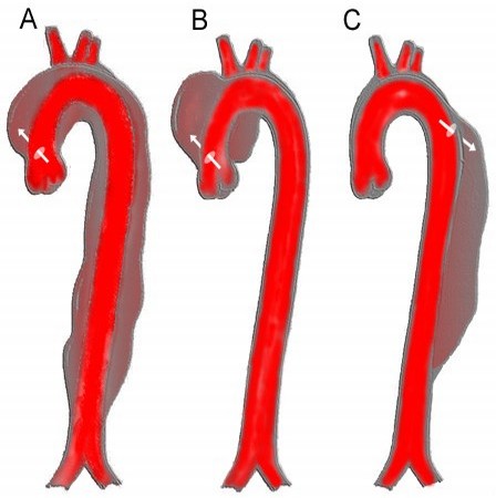
20. The types of aortic dissection are ascending (type A) dissection, which involves only the ascending or both the ascending and descending aorta, and descending dissection (type B), which involves only the descending aorta.


21. A solitary pulmonary nodule is < 3 cm and is discrete on chest radiograph. It is usually surrounded by lung parenchyma.
22. Mediastinal staging is indicated in patients with apparent or documented lung cancer who have (1) known lung cancer with mediastinal nodes > 1 cm accessible by cervical mediastinal exploration, as assessed by CT scan; (2) adenocarcinoma of the lung and multiple mediastinal lymph nodes < 1 cm; (3) central or large (> 5 cm) lung cancers with mediastinal lymph nodes < 1 cm; and (4) lung cancer with risk of thoracotomy and lung resection.
23. The most common causes of aortic stenosis are now congenital anomalies and calcific (degenerative) disease.
24. In mitral regurgitation, the left ventricle ejects blood via two routes: (1) antegrade, through the aortic valve, or (2) retrograde, through the mitral valve. The amount of each stroke volume ejected retrograde into the left atrium is the regurgitant fraction. To compensate for the regurgitant fraction, the left ventricle must increase its total stroke volume. This ultimately produces volume overload of the left ventricle and leads to ventricular dysfunction.
25. The indications for CABG are (1) left main coronary artery stenosis; (2) three-vessel coronary artery disease (70% stenosis) with depressed left ventricular (LV) function or two-vessel coronary artery disease (CAD) with proximal left anterior descending (LAD) involvement; and (3) angina despite aggressive medical therapy.
26. Hibernating myocardium is improved by CABG. Myocardial hibernation refers to the reversible myocardial contractile function associated with a decrease in coronary flow in the setting of preserved myocardial viability. Some patients with global systolic dysfunction exhibit dramatic improvement in myocardial contractility after CABG.
27. The surgical treatment of ulcerative colitis is total colectomy with ileoanal pouch anastomosis.
28. Dieulafoy's ulcer is a gastric vascular malformation with an exposed submucosal artery, usually within 2-5 cm of the gastroesophageal junction. It presents with painless hematemesis, often massive.
29. The role of blind subtotal colectomy in the management of massive lower gastrointestinal bleeding is limited to a small group of patients in whom a specific bleeding source cannot be identified. The procedure is associated with a 16% mortality rate.
30. Colorectal polyps < 2 cm have a 2% risk of containing cancer, 2 cm polyps have a 10% risk, and polyps > 2 cm have a cancer risk of 40%. Sixty percent of villous polyps are > 2 cm, and 77% of tubular polyps are < 1 cm at the time of discovery.
31. Patients with colorectal cancer with lymph node involvement (Dukes' C) should receive chemotherapy postoperatively to treat micrometastases.
32. Goodsall's rule states the location of the internal opening of an anorectal fistula is based on the position of the external opening. An external opening posterior to a line drawn transversely across the perineum originates from an internal opening in the posterior midline. An external opening, anterior to this line, originates from the nearest anal crypt in a radial direction.
Incarcerated inguinal hernia: structures in the hernia sac still have a good blood supply but are stuck in the sac because of adhesions or a narrow neck of the hernia sac. Strangulated inguinal hernia: hernia structures have a compromised blood supply because of anatomic constriction at the neck of the hernia.


33. Incarcerated inguinal hernia: structures in the hernia sac still have a good blood supply but are stuck in the sac because of adhesions or a narrow neck of the hernia sac. Strangulated inguinal hernia: hernia structures have a compromised blood supply because of anatomic constriction at the neck of the hernia.
34. Chvostek's sign is spasm of the facial muscles caused by tapping the facial nerve trunk. Trousseau's sign is carpal spasm elicited by occlusion of the brachial artery for 3 minutes with a blood pressure cuff.
35. The two surgical options for Graves' disease are subtotal thyroidectomy or near-total thyroidectomy.
36. The only biochemical test that is routinely needed to identify patients with unsuspected hyperthyroidism is serum thyroid-stimulating hormone concentration.
37. The surgically correctable causes of hypertension are renovascular hypertension, pheochromocytoma, Cushing's syndrome, primary hyperaldosteronism, coarctation of the aorta, and unilateral renal parenchymal disease.
38. The "triple negative test" or "diagnostic triad" for diagnosing a palpable breast mass includes physical examination, breast imaging, and biopsy.
39. Chest wall radiation is indicated after mastectomy in patients with greater than 5 cm primary cancers, positive mastectomy margins, or more than four positive lymph nodes, all of which are associated with heightened locoregional recurrence rates.
40. Sentinel lymph nodes are the first stop for tumor cells metastasizing through lymphatics from the primary tumor.
41. The most common site of origin of subungual melanomas is the great toe. Amputation at or proximal to the metatarsal phalangeal joint and regional sentinel lymph node biopsy are advised by most authors.
42. Ramus marginalis mandibularis, the lowest branch of the nerve that innervates the depressor muscles of the lower lip, is the most commonly injured facial nerve branch during parotidectomy.
43. Waldeyer's ring is the mucosa of the posterior oropharynx covering a bed of lymphatic tissue that aggregates to form the palatine, lingual, pharyngeal, and tubal tonsils. These structures form a ring around the pharyngeal wall. This may be the site of primary or metastatic tumor.
44. A patient in whom the head and neck examination is completely normal but FNA of a cervical node reveals squamous cancer should have examination of the mouth, pharynx, larynx, esophagus, and tracheobronchial tree under anesthesia (triple endoscopy). If nothing is seen, blind biopsy of the nasopharynx, tonsils, base of tongue, and pyriform sinuses should be done at the same sitting.
45. The microorganisms implicated in atherosclerosis include Chlamydia pneumoniae, Helicobacter pylori, streptococci, and Bacillus typhosus.
46. The cumulative 10-year amputation rate for claudication is 10%.
47. The absolute reduction in risk of stroke is 6% over a 5-year period in asymptomatic patients with > 60% stenosis who undergo carotid endarterectomy plus aspirin versus patients treated with aspirin alone (5.1% versus 11%).
48. Abdominal aortic aneurysm's average expansion rate is 0.4 cm/year.
49. Heparin binds to antithrombin III, rendering it more active.
50. The patient with suspected intermittent claudication should initially be evaluated by obtaining ankle brachial index or segmental limb pressures at rest.
51. Shock is suboptimal consumption of O2 and excretion of CO2 at the cellular level.
52. Nitric oxide is synthesized in vascular endothelial cells by constitutive nitric oxide synthase and inducible NOS, using arginine as the substrate.
53. Saliva has the hightest potassium concentration (20 mEq), followed by gastric secretions (10 mEq), then pancreatic and duodenal secretions (5 mEq).
54. Basal caloric expenditure = 25 kcal/kg/day with a requirement of approximately 1 g protein/kg/day.
55. 6.25 g of protein contains 1 g of nitrogen.
56. Dextrose has 3.4 kcal/g, protein 4 kcal/g, fat 9 kcal/g (20% lipid solution delivers 2 kcal/mL).
57. Maximal glucose infusion rates in parenteral formulas should not exceed 5 mg/kg/min.
58. Refeeding syndrome occurs in moderately to severely malnourished patients (e.g., chronic alcoholism or anorexia nervosa) who, upon presentation with a large nutrient load, develop clinically significant decreases in serum phosphorus, potassium, calcium, and magnesium levels. Hyperglycemia is common secondary to blunted insulin secretion. ATP production is mitigated, and the classic presentation is respiratory failure.
59. Glutamine is the most common amino acid found in muscle and plasma. Levels decrease after surgery and physiologic stress. Glutamine serves as a substrate for rapidly replicating cells (interestingly, it is also the number one metabolic substrate for neoplastic cells), maintains the integrity and function of the intestinal barrier, and protects against free radical damage by maintaing GSH levels. Glutamine is unstable in IV form unless linked as a dipeptide.
60. Fever is caused by activated macrophages that release interleukin-1, tumor necrosis factor, and interferon in response to bacteria and endotoxin. The result is a resetting of the hypothalamic thermoregulatory center.
61. Cardiac output = heart rate x stroke volume; normal CO is 5-6 L/min.
62. SVR = [(MAP - CVP)/CO] x 80; normal SVR is 800-1200 dyne.sec/cm-5.
63. Hypovolemic shock: low CVP and PCWP, low CO and SVO2, high SVR.
64. Cardiogenic shock: high CVP and PCWP, low CO and SVO2, variable SVR.
65. Septic shock: low or normal CVP and PCWP, high CO initially, high SVO2, low SVR.
66. Kehr's sign is concurrent LUQ and left shoulder pain, indicating diaphragmatic irritation from a ruptured spleen or subdiaphragmatic abscess. Anatomically, the diaphragm and the back of the left shoulder enjoy parallel innervation.
67. Rebound tenderness implies peritoneal inflammation and irritation not simply abdominal tenderness.
68. The 5 Ws of post-operative fever are wound (infection), water (UTI), wind (atelectasis, pneumonia), walking (thrombophlebitis), and wonder drugs (drug fevers).
69. Cricothyroidotomy should not be performed in patients <> 80 mmHg; femoral (groin) pulse estimates SBP > 70 mmHg; and carotid (neck) pulse estimates SBP > 60 mmHg.
71. A general rule for crystalloid infusion to replace blood loss is a 3:1 ratio of isotonic crystalloid to blood.
72. Raccoon eyes (periorbital ecchymosis) and Battle's sign (mastoid ecchymosis) are clinical indicators of basilar skull fracture.
73. CPP = MAP - ICP. Some debate exists on the minimum allowable CPP, but consensus indicates that a cerebral perfusion pressure of 50-70 mmHg is necessary.
74. Violation of the platysma defines a penetrating neck wound.
75. Tension pneumothorax is air accumulation in the pleural space eliciting increased intrathoracic pressure and resulting in a kinking of the SVC and IVC that compromises venous return to heart.
76. The most common site of thoracic aortic injury in blunt trauma is just distal to the take-off of the left subclavian artery.
77. The most common manifestation of blunt myocardial injury is arrhythmia.
78. Indications for thoracotomy in a stable patient with hemothorax include an immediate tube thoracostomy output of > 1500 mL and ongoing bleeding of 250 mL/h for 4 consective hours.
79. Beck's triad is hypotension, distended neck veins, and muffled heart sounds.
80. The hepatic artery supplies approximately 30% of blood flow to the liver while the portal vein supplies the remaining 70%. The oxygen delivery, however, is similar for both at 50%.
81. The Pringle maneuver is a manual occlusion of the hepatoduodenal ligament to interrupt blood flow to the liver.
82. Splenectomy significantly decreases IgM levels.
83. 90% of trauma fatalities due to pelvic fractures are due to venous bleeding and bone oozing; only 10% of fatal pelvic bleeding from blunt trauma is arterial (most common site is superior gluteal artery).
84. Intraperitoneal bladder rupture from blunt trauma: operative management; extraperitoneal rupture: observant management.
85. Pseudoaneurysm is a disruption of the arterial wall leading to a pulsatile hematoma contained by fibrous connective tissue (but not all three arterial wall layers, which defines a true aneurysm).
86. The earliest sign of lower extremity compartment syndrome is neurologic in the distribution of the peroneal nerve with numbness in the first dorsal webspace and weak dorsiflexion.
87. Posterior knee dislocations are associated with popliteal artery injuries and are an indication for angiography.
88. Management of suspected navicular fracture despite negative radiography is short-arm cast and repeat x-ray in 2 weeks; at high risk for avascular necrosis.
89. Parkland formula: lactated Ringer's at 4 mL/kg x %TBSA (second- and third-degree only) of burn. Infuse 50% of volume in first 8 hours and the remaining 50% over the subsequent 16 hours.
90. The metabolic rate peaks at 2.5 times the basal metabolic rate in severe burns > 50% TBSA.
91. Gallstones and alcohol abuse are the two main causes of acute pancreatitis.
92. Alcohol abuse accounts for 75% of cases of chronic pancreatitis.
93. Isolated gastric varices and hypersplenism indicate splenic vein thrombosis and are an indication for splenectomy.
94. The treatment for gallstone pancreatitis is cholecystectomy and intraoperative cholangiogram during the same hospital stay once the pancreatitis has subsided.
95. Proton pump inhibitors irreversibly inhibit the parietal cell hydrogen ion pump.
96. Definitive treatment of alkaline reflux gastritis after a Billroth II includes a Roux-en-Y gastro-jejunostomy from a 40-cm efferent jejunal limb.
97. Cushing's ulcer is a stress ulcer found in critically ill patients with central nervous system injury. It is typically single and deep, with a tendency to perforate.
98. Curling's ulcer is a stress ulcer found in critically ill patients with burn injuries.
99. Marginal ulcer is an ulcer found near the margin of gastroenteric anastomosis, usually on the small bowel side.
100. The most common cause of small bowel obstructions is adhesive disease; the second most common cause is hernias.


read more about“Top 100 Surgical Secrets”
TUMOURS OF THE PANCREAS
6/02/2009 09:59:00 PM | Author: fadzly
Let's talk about Pancreatic tumour. There are many types of pancreatic tumour. Although ductal carcinoma of the pancreas is one of the most lethal of all gastrointestinal tumours, 10–15% of tumours of the pancreas (including those of the periampullary region) are not of the same histological type as ductal carcinoma and have a much better prognosis. This group of tumours includes ampullary carcinoma, islet cell tumours of the pancreas and cystic tumours of the pancreas. A tumour of the pancreas should, therefore, not be assumed to carry a poor prognosis. Ok?

Ductal adenocarcinoma of the pancreas is highly malignant and has usually metastasised by the time of diagnosis. Smoking is the only recognised aetiological factor and the disease is uncommon under the age of 40. The disease occurs in the head of the pancreas in 80% of cases. It presents with obstructive jaundice in 70% of cases but may also present with severe upper abdominal pain (which may radiate into the back), weight loss, anorexia, malaise, or rarely, thrombophlebitis migrans.

On examination, there may be cervical lymphadenopathy, an abdominal mass, hepatomegaly or ascites. The gallbladder may be palpable. Courvoisier’s law states that if, in the presence of jaundice, the gallbladder is palpable, then the cause is unlikely to be gallstones. Investigations include basic blood tests and specific diagnosis by ultrasound, CT or MRI. The ERCP may be helpful too, and may also allow therapeutic manoeuvres such as stent insertion (see section on obstructive jaundice).

Most tumours are treated palliatively by insertion of a biliary stent or bypass surgery. Resectional surgery is suitable for 15–20% of patients with a ductal carcinoma but 50–75% of patients with ampullary carcinoma, islet cell tumours or cystic tumours of the pancreas. Five-year survival following curative resection in ductal carcinoma is 10–15% although resection is also associated with good relief of symptoms. For those with ampullary tumours, islet cell tumours or cystic tumours, 5 year survival of 40% can be anticipated.

For tumours of the head of the pancreas or peri-ampullary region, pancreatico-duodenectomy (Whipple’s operation) is the operation of choice. The pylorus-preserving operation is the operation of choice. The mortality rate of pancreatico-duodenectomy is less than 5%.


Whipple’s procedure (partial pancreatoduodenectomy) for carcinoma of the
head of pancreas (shaded).

read more about“TUMOURS OF THE PANCREAS”
SURGERY: Biliary Tract
5/30/2009 08:27:00 PM | Author: fadzly
Topic today will be about Biliary tract


GALLSTONES

Gallstones are very common topic and are therefore frequently asked in the exams. Majority of them are asymptomatic and which require no treatment. However, they cause a wide range of clinical problems, depending on their position. They are either made up of cholesterol or pigment (bilirubin breakdown products) or a mixture of the two. Most (75%) gallstones are predominantly cholesterol. Pure pigment stones are rare (<10%) Ninety per cent of gallstones are radiolucent, i.e. they do not show on a plain X-ray (unlike renal calculi, of which 90% are radio-opaque).

Predisposing factors to gallstone formaton include:
1. female
2. sex (three times more common)
3. obesity,
4. age (10% of <50-year-olds have gallstones, and 30% of <70-year-olds),
5. Haemolytic anemia
6. Hyperlipidaemias
7. Crohn’s disease.


Some people appear to have a tendency to form gallstones and are said to have ‘lithogenic bile’. People often refer to the typical gallstone patient as 4F which is fat, female, fertile and forty. Ok? So remember that 4F if u cant remember the risk factors

Complications of gallstones can be divided into 3:
1. In the gallbladder
  • Chronic cholecystitis
  • Biliary colic
  • Acute cholecystitis
  • Empyema
  • Biliary peritonitis
  • Abscess
  • Mucocele
  • Carcinoma of gallbladder
2. In the common bile duct
  • Obstructive jaundice
  • Cholangitis
  • Pancreatitis
3. In the gut
  • Gallstone ileus

BILIARY COLIC

Biliary colic is the pain that associated with irritation of the viscera secondary to cholecystitis and gallstones or in a specific terms caused by gallbladder muscle spasms against a stone stuck in the neck of the gallbladder (Hartmann’s pouch) or the cystic duct. It may account for some of the symptoms for chronic cholecystitis. Unlike intestinal colic, the pain was continuous and not in waves. It is usually felt in the epigastrium or right upper quadrant and may radiate around both costal margins and into the back. The pain can be extremely severe and patients may be sweaty, pale and tachycardic because of it. They may also feel nauseated or vomit. They will usually be unable to get comfortable and will prefer to writhe around rather than stay still. Attacks usually last less than six hours and examination is usually otherwise normal. Differential diagnoses include other causes of severe upper abdominal pain, such as perforated peptic ulcer, pancreatitis, ruptured aneurysm, etc. Management involves giving analgesia, investigation to confirm gallstones (ultrasound) and subsequent cholecystectomy in most cases.

ACUTE CHOLECYSTITIS

History

At early stage acute cholecystitis may appear to be biliary colic, and indeed many attacks of acute cholecystitis probably start with biliary colic! The exact mechanisms of acute cholescystitis are poorly understood but they said that it are caused by chemical inflammation within an obstructed gallbladder. Bacterial infection probably is a secondary event in about one-third of cases and these may be the ones most likely to develop complications.

Patients will typically have symptoms of severe right upper quadrant or epigastric pain. Like biliary colic, this may radiate around the costal margins or into the back. However unlike biliary colic, patients will prefer to lie still and take shallow breaths (this is now a form of local peritonitis, not colic). They will usually have a temperature and tachycardia, and may also have nausea and vomiting.

Physical examination

Murphy’s sign may be positive and is often asked about in short case. It is elicited by pressing in the right upper quadrant under the costal margin. The patient is then asked to breathe in, and winces or gasps with pain as the gallbladder moves down and hits the examiner’s hand. The test should also be performed in the left upper quadrant to exclude nonspecific reactions due to other pathology.

A mass may be present in the right upper quadrant, but if so this is not usually the gallbladder itself but rather a ‘phlegmon’ (i.e. inflamed andadherent omentum and bowel around the gallbladder).

Investigation

The most important confirmatory test is an ultrasound scan. US confirm gallstones, showing thickening and oedema of the gallbladder wall and localise the tender spot to the gallbladder itself. It can also exclude dilatation of the common bile duct and other pathology, such as liver masses.

Image through the long axis of the GB (GB) demonstrates the gallbladder neck (red arrow). GB wall thickness is measured between the gallbladder lumen and the hepatic parenchyma (red arrowheads) with normal thickness <3cm

Only very occasionally is a HIDA scan used to help confirm or exclude cholecystitis. The principle of this test is that HIDA (a radioisotope) is taken up by the liver and excreted into the bile. If the cystic duct is patent, it will fill the gallbladder effectively, excluding cholecystitis.

Management

The treatment of acute cholecystitis is initial resuscitation with intravenous
fluids and antibiotics. The patient will normally be kept nil by mouth or on sips of clear fluids, and initial investigations will be arranged, including basic blood tests such as an FBC (usually the white cell count is raised), U & Es, LFTs and amylase (as acute pancreatitis may be a differential diagnosis).

With conservative treatment approximately 80–90% of cases of acute cholecystitis will settle over the next 24–48 h (i.e. the pain settles, the temperature falls and the patient’s abdomen becomes nontender). In about 10% of cases there will not be a prompt resolution of symptoms and signs, and in these cases surgery is usually advised.

Particularly worrying signs are increasing temperature, tachycardia and the onset of increasing tenderness or the signs of peritonitis. These may indicate infarction of the gallbladder (gangrenous cholecystitis) or perforation, which may produce either a local collection or generalised peritonitis. A gallbladder full of pus (empyema of the gallbladder) usually leads to an unwell patient with the signs of sepsis (fever, tachycardia, hypotension, etc.) as well as pain, and tenderness in the right upper quadrant.

More controversial is the question of what to do with patients who do not absolutely require early surgery. Although conventional management is to allow the acute episode to settle down and to readmit the patient for elective cholecystectomy 6–8 weeks later, many surgeons now prefer cholecystectomy in the acute phase. This allows patients to recover quicker and to be spared further episodes of pain. Laparoscopic cholecystectomy can now be carried out in the acute phase by experienced surgeons.


CHRONIC CHOLECYSTITIS

Chronic cholecystitis is a term used to describe symptoms of upper abdominal pain, indigestion, bloating, burping, nausea and occasional vomiting. Sometimes this symptom complex is called flatulent dyspepsia. The patient may describe the symptoms as being precipitated by fatty food (fats stimulate the release of cholecystokinin, which causes gallbladder contractions). There is usually nothing to find on physical examination.

The main differential diagnoses include
1. peptic ulceration
2. hiatus hernia
3. irritable bowel syndrome.

Because gallstones are so common, it's important not to automatically discribe such symptoms to them simply because that gallstones are present on an US scan. A missed peptic ulcer or irritable bowel syndrome will obviously not be helped by unnecessary cholecystectomy! And the patient will continue to get those symptoms. If the symptoms are thought to be arising from the gallbladder and are significant, then the treatment is cholecystectomy, usually laparoscopic.

Attempting to dissolve gallstones using 'bile salt therapy' might be possible for patients with small, noncalcified stones. It also reserved for those who refuse or unfit for surgery. However bear in mind that this treatment is not very successful.

CHOLANGITIS

Cholangitis is a condition where there is infection within the biliary tract and it is rare unless there is associated obstruction. This is a demonstration of the surgical principle that obstructed tubes tend to get infected, i.e. appendicitis, pyelonephritis, etc. Cholangitis is clinically manifested by Charcot’s triad of pain, jaundice and rigors (rigors means involuntary shaking in association with pyrexia). It requires prompt diagnosis and treatment, otherwise it can have a high mortality. Treatment consists of resuscitaton with fluids and the administration of intravenous antibiotics. If resolution is not rapid, then attempts to produce biliary drainage, endoscopically, radiologically or surgically, are required.

That's all for today! Happy revising!


read more about“SURGERY: Biliary Tract”
Warning: GRAPHIC MEDICAL IMAGES BELOW
5/19/2009 05:37:00 AM | Author: fadzly
I was thinking of clerking a breast cancer patient today. It's common for a medical student except me (i mean i try to be common today, sigh so delusion of me), before ever clerking a patient, you need to be at least have a basic knowledge of the disease and i've managed to get into the web and searching for the relevant articles and some pictures of breast cancer. In the middle of surfing the net, I was totally shock of what I’ve found. Guys, before u go any futher, I really need to warned you, if you not prepare for anything below or if you’re below 18 years old, don’t ever think of scrolling down and CLOSE THIS WINDOW IMMEDIATELY! You might not having breakfast today or even worse your lunch. I'll not be responsible for what happen to you after you watch the picture below.
It started as an email sending around in the web, a medical case of a female anthropologist named Susan McKinley who failed to seek treatment for a rash and wound up with an infestation of larvae in her breast. OMG. Larvae in her breast???

The mail was like this

Fwd: Wash before Wearing!
It is horrible. Guys tell your wife, sisters, girlfriends, and girl cousins wash bra before wearing.
ALL PLEASE WASH ALL BRAS, UNDERWEAR WHEN YOU BUY BEFORE WEARING THEM. WE DO NOT KNOW WHAT PARASITE IS IN OUR CLOTHES WHEN WE BUY THEM. FORWARD TO EVERYBODY YOU KNOW. LET ME FORE WARN YOU THIS IS SO SQUIMISH, I FEEL LIKE SOMETHING IS CRAWLING ALL OVER ME EVEN AS I SEND THIS TO YOU. BE AWARE. IT IS SO GROTESQUE. PLEASE WASH YOUR UNDERWEAR BEFORE WEARING. PREFERABLY IN BOILING HOT WATER.
This is not for the weak; I have never seen anything like this. Read the article first before looking at the picture and film. This looks horrible. Oh my God!!!!!!! Ladies this could happen to you and Guys this could happen to your wife, girlfriend, partner so please BEWARE,and also warn others.
It has been reported that this is happening in Zimbabwe as well, please make sure you iron your undergarments before you wear them and make sure that your clothes are ironed when they are dry and not damp. The picture is horrible but I felt that I should share with you. After anthropologist Susan McKinley came back home from an expedition in South America, she noticed a very strange rash on her left breast. Nobody knew what it was and she quickly dismissed it believing that the holes would leave in time. Upon her return she decided to see a doctor after she started developing intense pains. The doctor, not knowing the exact severity of the disease, gave her antibiotics and special creams. As time lapsed the pain did not subside and her left breast became more inflamed and started to bleed.
She decided to bandage her sores however as Susan's pain grew more intense she decided to seek help from a more certified doctor. Dr. Lynch could not diagnose the infection and told Susan to seek the aid of one of his colleagues who specialized in dermatology whom was sadly on vacation. She waited for two weeks and finally was able to react the dermatologist.
Sadly,a life changing event was about to unfold during her appointment.
To Miss McKinley's surprise, after she removed the bandages, they found larva growing and squirming within the pores and sores of her breast. Sometimes these wicked creatures would all together simultaneously move around into different crevices.
What she didn't know was that the holes were in fact, deeper than she had originally thought for these larvae were feeding off the fat, tissue, and even milk canals of her bosom.

Picture and Video


Comment
The picture and the video above, definitely, u can see without even taking a photofake diploma, u can see the difference between them. The top one might have been photoshop. Of coz is a fake. How come the breast so clean on the outerpart, and there is no sign of any inflammation going on there. Later i found this picha. Ah-ha! They fake by photoshopped a lotus seed pod onto a breast.

The video although, was not a fake, but seems to me, the video was not referring to Susan McKinley (I mean white people is not a black people isn’t it?).
Hey man, at first I was just trying to look for complication of Breast Cancer. This larvae in the breast is a true medical condition where the terms “Myiasis of the breast”. There even one case in UMMC I couldn’t remember when, but it was a complication of breast cancer usually at the end stage (stage 4th) cancer. I never thought that this thing might occur but one of my friends said that larvae infestations of the breast, though extremely rare, can actually occur without having any of those co-morbidities!
Then there is one note saying to all women to wash their bra after buying it. I was thinking, whether the video who I think it might comes from an African woman wearing bra or not.. LOLZ..

For me, I would like to suggest to all ladies or even some men, don't wait until your breast become like this then it's too late for treatment. Practice self breast examination, so that you will learn your breast better. It's a simple screening, yet it saving life's.

It’s 6.30 a.m now. Better get ready for breakfast and go to the wards. See ya later…
read more about“Warning: GRAPHIC MEDICAL IMAGES BELOW”